
Being an antibiotic-resistant cause of life-threatening pneunomia, it is no wonder that Pseudomonas aeruginosa is such a notorious and feared pathogen. But even so, much remains unknown about this bacterium. How does it succeed in deeply penetrating lung tissue? By studying the infection of lung organoids, Swiss researchers revealed the bacterium’s Trojan horse-stategy.
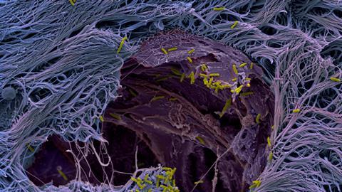
High on the World Health Organization’s list of bacterial priority pathogens we find Pseudomonas aeruginosa, the little yellow rods in this picture that are working their way into human lung tissue (purple). The picture is a SEM (Scanning Electron Microscope) image and the lung tissue is an organoid, cultured from human stem cells.
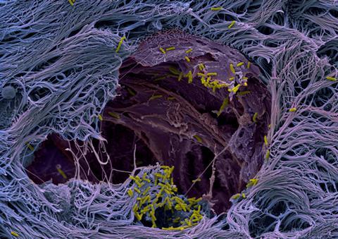
The team of Urs Jenal at the Unversity of Basel (Switzerland) used these ‘mini-lungs’ to understand how P. aeruginosa manages to penetrate the protective layer of mucus on the lung epithelium. It turns out that the bacterium deploys a Trojan horse-strategy: P. aeruginosa secretes compounds that specifically target the so-called goblet cells. These cells form the outer layer that acts as a protective barrier of the underlying tissue.
Next, the bacteria colonize the weakened goblet cells and start multiplying untill the cells burst. This creates ruptures in the protective barrier, enabling P. aeruginosa to easily penetrate deeper into the tissue. According to Jenal, this study demonstrates the importance of using human organoids to really grasp how pathogens operate, which is a crucia step towards the development of effective treatments.
Swart, A.L., Laventie, BJ., Sütterlin, R. et al, Pseudomonas aeruginosa breaches respiratory epithelia through goblet cell invasion in a microtissue model, Nature Microbiology (2024), doi:10.1038/s41564-024-01718-6


Nog geen opmerkingen