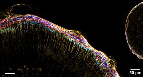
Amsterdam-based Confocal.nl has received a €5 million investment to make its live cell imaging more accessible. As a foretaste of what is possible, they have produced this stunning image of a mouse ear.
Microscopy has come a long way since the lenses of Antoni van Leeuwenhoek. A spin-off from the University of Amsterdam, Confocal.nl, uses Re-scan confocal technology to capture live images of biological cells. This image shows the inner ear (cochlea) of a mouse.
What makes the company unique is that it uses minimal laser light at very high resolution, allowing you to observe living cells longer and better. ‘Traditional microscopes use intense laser light, which causes cells to behave differently, fade or die more quickly. As a result, the ability to study living cells is limited. Our technology overcomes this and allows scientists to study processes in living cells for longer and more accurately, with more reproducible results (…),’ said CEO Pim Vos in a press release. With the €5 million investment, the company aims to raise awareness of its technology and make it more accessible to scientists and companies that want to use its service.
Do you have nice pictures from your research too? Send them to redactie@kncv.nl and we might feature you in Photo Chemistry!


Nog geen opmerkingen