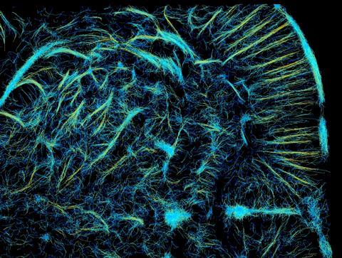
We know ultrasounds as grainy grey pictures, but with the latest imaging techniques, they can also look like the picture above. Eindhoven researchers visualised the vascular structure of a rat brain using advanced ultrasound.
Many arteries and capillaries are so small that you can’t see them with conventional ultrasound. However, super-resolution ultrasound allows you to see these vessels in high resolution. This image shows the vascular structure of the brain of a living rat. To create this image, researchers injected so-called contrast bubbles, about the size of a red blood cell, into the blood vessels. These contrast bubbles reflect sound waves very strongly, allowing you to localise them very precisely. The researchers tracked the individual bubbles over several frames, after which they were able to plot the entire vascular structure. The intensity of the colour shows where blood flow is best.
Tristan Stevens and his team from TU Eindhoven and Philips won second prize with their technique at the International Ultrasound Symposium in Venice (IUS2022).

Sources:
Picture:
Stevens, Tristan S.W., et al (2022). “A Hybrid Deep Learning Pipeline for Improved Ultrasound Localization Microscopy.” 2022 IEEE International Ultrasonics Symposium (IUS). IEEE, 2022. http://dx.doi.org/10.1109/IUS54386.2022.9958562
Data:
Lerendegui, M., Riemer, K., Wang, B., Dunsby, C., & Tang, M. (2022). BUbble Flow Field: a Simulation Framework for Evaluating Ultrasound Localization Microscopy Algorithms. arXiv. https://doi.org/10.48550/arXiv.2211.00754


Nog geen opmerkingen